Tracheostomy Tube
A tracheostomy tube, or trach tube, is a hollow tube inserted through a surgical opening in the neck into the trachea (windpipe) to create an artificial airway. This procedure, called a tracheotomy, allows air and oxygen to reach the lungs, especially when breathing through the mouth and nose is difficult or obstructed.
Minimum Quantity Order: 200 Unit
Price: Negotiable
Product Enquiry Form
Sharing is caring:-
Description
A tracheostomy tube, or trach tube, is a hollow tube inserted through a surgical opening in the neck into the trachea (windpipe) to create an artificial airway. This procedure, called a tracheotomy, allows air and oxygen to reach the lungs, especially when breathing through the mouth and nose is difficult or obstructed.
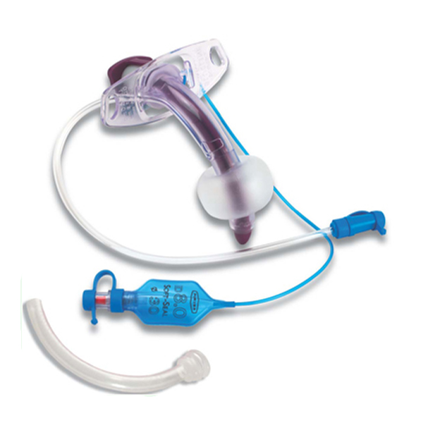
Components of a tracheostomy tube
Tracheostomy tubes have several standard components:
- Outer Cannula: The main shaft of the tube inserted into the trachea.
- Inner Cannula: A disposable or reusable tube that slides inside the outer cannula, facilitating cleaning and preventing obstruction by mucus build-up. Single-cannula tubes don’t have this component.
- Flange (Neck Plate): The part that rests against the neck and has holes for securing the tracheostomy tube with ties. The flange often provides information about the tube’s size, diameter, brand, and cuff type.
- Cuff: A balloon-like feature near the end of the outer cannula. Cuffed tubes are used to seal the airway, preventing aspiration (inhaling food or liquid) and ensuring effective mechanical ventilation. Low-pressure, high-volume cuffs are more common and are typically filled with air. Low-volume, high-pressure cuffs, like Bivona TTS, are usually single lumen and are filled with sterile water. Foam-filled cuffs are sometimes used in cases of tracheal injury or tracheomalacia.
- Pilot Balloon and Inflation Line: These are present on cuffed tubes and indicate the cuff’s inflation status. The inflation line leads from the cuff to the pilot balloon, allowing inflation and deflation of the cuff with a syringe attached to the luer valve.
- Obturator: A solid, blunt-tipped device used during insertion to guide the tracheostomy tube and prevent tissue damage. It is removed after insertion.
- Tracheostomy Tube Tie: Secures the tube to the neck through the flange.
Types of tracheostomy tubes:
In addition to cuffed and uncuffed tubes, other types exist:
- Fenestrated Tubes: These have holes (fenestrations) in the outer cannula to allow airflow through the vocal cords, enabling speech.
- Single Lumen Tubes: Consist only of an outer cannula without an inner cannula. They are typically used for pediatric patients due to their smaller airways.Double Lumen Tubes: Also known as dual cannula tubes, they have both an inner and outer cannula.
Tracheostomy tube sizes:
Tracheostomy tube sizes are not standardized across manufacturers, so it’s important to consider the tube’s inner diameter (ID), outer diameter (OD), and length, rather than relying solely on the manufacturer’s size label. The length and diameter of the trachea are roughly proportional to the individual’s size.
Materials used for tracheostomy tubes
Most modern tracheostomy tubes are made from medical-grade polyvinyl chloride (PVC), polyurethane, silicone, or a combination of these materials. In the past, they were made of rubber, which could be reused after sterilization. Metal tubes, like the Jackson tube, are also available.
Tracheostomy tube insertion and removal
A tracheotomy is a surgical procedure to create the opening, into which a tracheostomy tube is inserted.
Insertion (Tracheotomy):
- This can be done surgically in an operating room or using a minimally invasive percutaneous approach.
- During an open surgical tracheotomy, the surgeon makes an incision in the neck to expose the windpipe and then creates an opening to insert the tracheostomy tube.
- For a percutaneous tracheostomy, a needle and then a catheter are used to guide and widen the opening for the tube.
- The tube is then secured with ties around the neck and may also be temporarily stitched in place.
Removal (Decannulation):
- When a tracheostomy is no longer needed, the tube can be removed without surgery in a doctor’s office.
- The tube is removed, and the opening is covered with a dressing.
- The tracheostomy will usually close on its own within a few weeks. If it doesn’t, a minor procedure may be needed to close it.
Caring for a tracheostomy tube:
Proper care is crucial to prevent complications like infection and obstruction.
- Suctioning: Regularly suctioning the tracheostomy tube removes mucus and secretions, maintaining a clear airway.
- Cleaning the Inner Cannula: If a dual cannula tube is used, the inner cannula should be cleaned or replaced regularly to prevent obstruction.
- Cleaning the Stoma: The skin around the tracheostomy site should be cleaned daily to prevent infection and irritation.
- Tracheostomy Ties: These should be changed regularly to maintain hygiene and ensure the tube is securely in place.
- Humidification: Because the upper airway is bypassed, the air entering the trachea may be dry. Using a humidifier or a heat and moisture exchanger (HME) helps to moisten the air and thin secretions.
- Speaking Valves: These can be attached to the tracheostomy tube to allow air to pass through the vocal cords, enabling speech.
Complications associated with tracheostomy tubes
Potential complications can occur during or after the procedure, or during long-term use.
Early Complications:
- Bleeding
- Damage to the windpipe, thyroid, or nerves
- Tube misplacement
- Subcutaneous emphysema (air trapped under the skin)
- Pneumothorax (air between the chest wall and lungs)
Late Complications:
- Tracheostomy tube blockage or displacement
- Tracheal damage, scarring, or narrowing (tracheal stenosis)
- Tracheoesophageal fistula (connection between the windpipe and food pipe)
- Infection
- Tracheomalacia (weakening of the tracheal wall)
- Granulation tissue